How Does Medical Imaging Analysis Revolutionize Diagnostic Procedures?
By playing a vital role in the diagnostic process, this medical imaging analysis has completely reshaped the way physicians look through the complex body for issues and cures. The new renaissance in medicine has emerged through the use of more advanced technologies and intelligent algorithms, it is now possible to explore areas that were not available earlier for instance, identifying abnormalities, accuracy of treatment, or guidance during surgical procedures. Let us explore the critical role of analyzing medical imaging and diagnostics in redefining diagnostic possibilities as they observe its use, appreciation, and projected future applications.
The Evolution of Medical Imaging
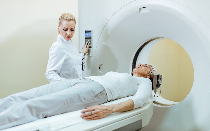
Medical imaging experienced a phenomenal change where there was the transition from the plain X-ray method to complex imaging tools of the type constituting computed tomography, magnetic resonance imaging, ultrasound, and positron emission tomography. Each modality has its strength and it allows the physicians to look at different parts of the body, its function, and signs of diseases using a never been done accuracy and clarity.
Enhanced Diagnostic Accuracy
One of the main advantages of medical imaging analysis is its ability to build up precise diagnostic correctness. Modern diagnostic methods give physicians the ability to localize abnormalities in early stages, assess disease developments, and track the response of the treatment method with admirable accuracy. Likewise, MRI, with its high soft tissue contrasting property, is a necessity for the visualization of brain structures, musculoskeletal system structures, and oncological imaging which others cannot obtain.
Early Detection and Intervention
Evaluation of medical imaging ranks among the key factors affecting early-stage disease detection and intervention allowing medical professionals to more preferably identify pathologies at the nascent stage when the prognosis is favorable. An example that proposes this method is mammography, an imaging technique that runs on the basis of X-rays. This helps detect breast cancer in its initial phases, which results in an early intervention that consequently improves patients’ treatment outcomes.
Benefits of Early Intervention
The early intervention phase is concerned with the implementation of treatment forms or preventative measures booming immediately in reaction to the positive findings from the early detection phase. There are many clear benefits to early intervention: improving treatment outcomes, slowing the progression of the illness, and enhancing patient well-being are just a few. Clinical cancer studies have been able to illustrate, for example, the ability for localized tumor resection to be done before metastasis, therefore, the chances of cure can be increased.
Emerging Technologies and Innovations
Medical imaging technologies have benefited from improved automation and standardization which is enabled by artificial intelligence (AI) and machine learning offering the promise of better diagnostics and early intervention. AI algorithms can process gigabytes of medical images from several patients and find out patterns related to the illnesses at an earlier stage therefore accurate diagnosis is done. To complement that, molecular imaging methods, such as PET/CT fusion imaging offer an opportunity to see biological processes at the molecular level which becomes another component of early detection.
Guiding Therapeutic Interventions
Besides diagnosis, medical imaging analysis acts in many situations as a tool to guide therapeutic interventions, neither in the field of interventional radiology nor in the field of image-guided surgery. By putting imaging on top of the real-time guidance systems for clinicians, it is possible to create effortless guidance for movements that can be able to target the lesions, give the therapy, and generally do minimally invasive procedures with unmatchable accuracy and safety.
Challenges and Future Directions
Despite numerous advantages of medical image analysis, the field is troubled by three significant issues, the first of them being data standardization, interoperability, and AI ethical concerns. These difficulties can be solved by bringing members of cross-disciplinary groups such the radiologists, imaging scientists, computer scientists, and health policy makers together.
Quantitative Analysis and Predictive Modeling
Innovations in medical image interpretation give grounds for numerical analysis and increased forecasting powers that save data from images for model development. Through computational algorithms and machine learning approaches, medical images are able to be quantified by their biomarkers, prognostic indicators, and thus treatment response forecasting models.
Personalized Medicine and Precision Imaging
The recent discovery of the power of the correlation of medical imaging analysis with genomic data, clinical parameters, and patient-specific data provides the foundation for personalized medicine and precision imaging. Through the application of targeted diagnostic and treatment modalities to individual patient-specific peculiarities, clinicians can obtain the best possible outcomes, diminish potential risks, and keep patients happy.
Applications of Precision Imaging
The armamentarium of precision imaging covers the broad domain of many branches of medicine such as oncology, cardiology, neuroscience, and rheumatology. In the field of oncology, precision imaging modalities having either functional MRI, diffusion-weighted imaging or PET/CT promote clinician to characterize the tumors, appraise their aggressiveness, and deliver targeted therapies by the molecular signatures.
Advancements in Radiomics and Radiogenomics
The last few years have witnessed rapid expansion in radiomics and radiogenomics which are now playing crucial roles in guiding precision imaging and obtaining quantitative imaging attributes and the linkages with genomic information for effective disease diagnoses and treatment selection. Through the detection and measurement of radionic features present in medical images, investigators can discover imaging biomarkers, predict the efficacy of treatment, and categorize patients according to risk categories in order to make the treatment customized.
Challenges and Considerations
Although it opens new prospects for personalized medicine and precision imaging, data normalization, interoperability, privacy issues, and regulatory barriers either already standing or about to emerge are very significant thorns on the sides of the precision medicine community. Such a procedure also means a big challenge related to advanced data from various sources which require the use of sophisticated computing algorithms, a solid infrastructure as well as interdisciplinary cooperation among clinicians, imaging scientists, bioinformaticians, and healthcare policymakers.
Conclusion
In conclusion, medical imaging analysis represents a cornerstone of modern diagnostics, offering unparalleled capabilities for early disease detection, treatment guidance, and personalized medicine. As technology continues to evolve and novel techniques emerge, the future of medical imaging analysis holds immense promise for revolutionizing diagnostic procedures and improving patient outcomes.


